Molar hypomineralization (MH), also known as “Molar-Incisor Hypomineralization” or “chalky teeth,” is a widespread condition that affects 19% of children worldwide.
Most often, the first adult molar erupts with spots of chalky enamel, but other adult or baby teeth can be affected. The “chalky” spots consist of enamel that is softer because it contains excess protein that prevents the full mineralization, making the enamel more prone to tooth decay.
MH can cause dental pain, and other dental problems. Its cause is unknown, and treatment strategies often do not work well in the soft enamel.
However, a recent study on enamel mineralization in pigs’ teeth provides new insight that could inform future directions in human MH treatment.
ADA Forsyth scientists examined whether the excess protein can be removed from enamel naturally in pig teeth. Proteins in developing teeth serve as ‘scaffolding’ that initially guides the growth of enamel crystals. To complete the mineralization, the proteins are removed and replaced by mineral, which fill in pores and harden tooth enamel.
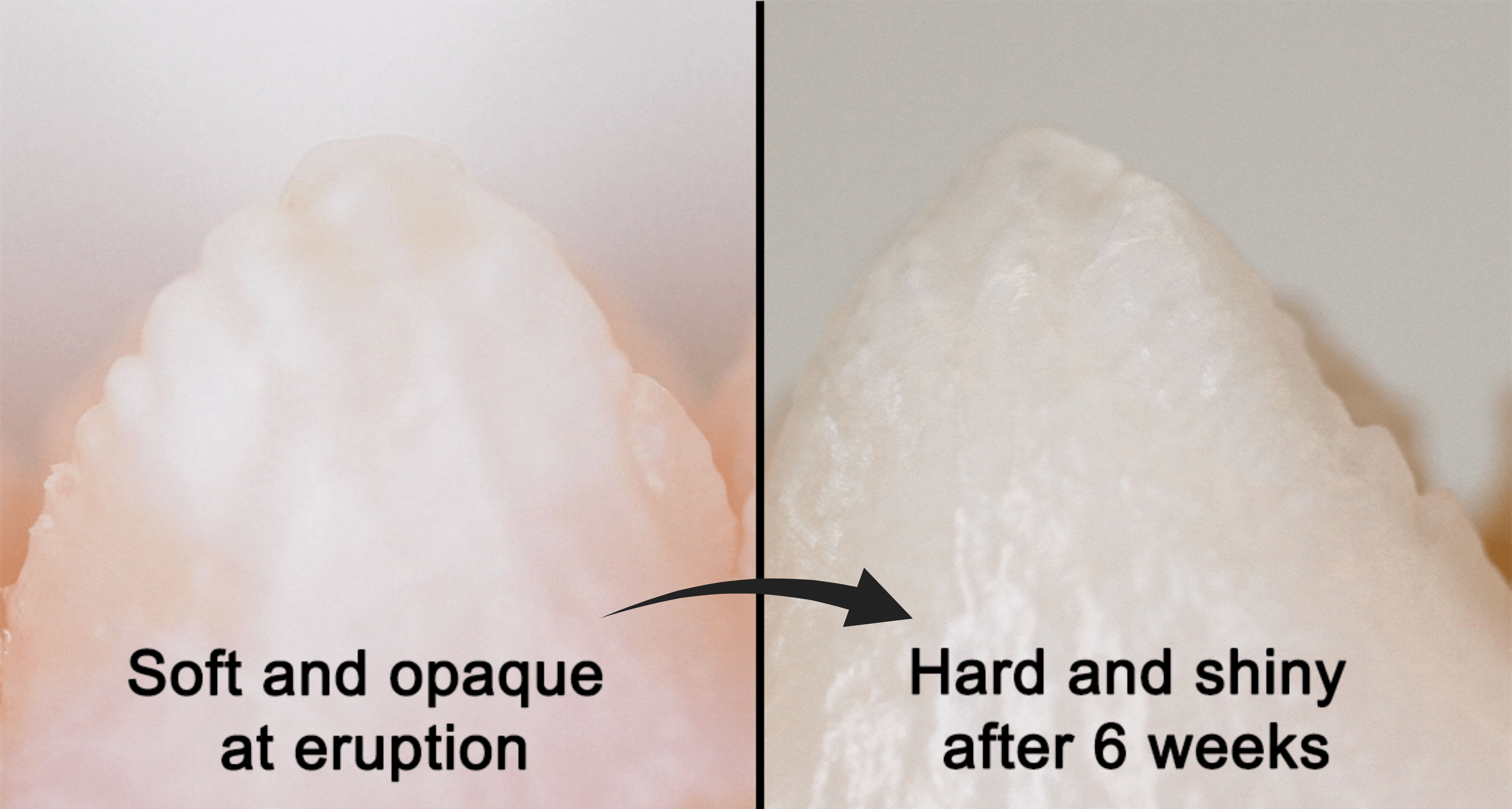
The process of hardening enamel in pig teeth is interesting to scientists because pig teeth erupt with soft enamel and harden naturally and quickly.
In contrast, when human teeth erupt with soft enamel, as in MH, this natural, fast mineralization does not occur. The AFI researchers looked at the fast mineralization in pig teeth to observe how a similar process could potentially be engineered in human teeth.
The study’s key finding showed that proteins in pig enamel decreased rapidly after teeth erupt and enter the oral environment. Enzymes from saliva likely facilitate the removal of proteins from enamel. Researchers identified enzymes which are likely to play a role in removing proteins and fortifying enamel.
“There might be a mechanism through which pigs remove those proteins, and the No. 1 pathway that we hypothesize is through saliva,” said ADA Forsyth postdoctoral researcher Dr. Hakan Karaaslan, D.D.S., the first author on the study, “Posteruptive Loss of Proteins in Porcine Enamel,” which was published in the Journal of Dental Research.
Pig teeth are similar in size and shape to human teeth, making them useful models for human biology in scientific research, Dr. Karaaslan said. They examined porcine teeth at three points in time: two weeks, four weeks and eight weeks after eruption.
Hypomineralization in humans can be treated with fluoride or other restorative treatments such as fillings, but patients would benefit greatly from a yet-to-be-developed alternative treatment which specifically removes the excess proteins and hardens enamel, avoiding the need for fillings.
Present treatments for hypomineralized enamel in humans use chemicals which can damage other organic parts of teeth.
“If we can have the same transformation in human patients with MH as displayed in pig teeth, that would improve the treatment outcomes of these patients in general,” Karaaslan said.
This work was supported by NIH grants R01DE025865 (to F.B.B), R21DE029903 (to F.B.B) and R90DE027638 (to H.K.).

