The ADA Forsyth Institute’s Core Facilities and Services provides specialized equipment, training and expertise on a fee-for-service basis to researchers from external institutions. AFI’s state-of-the-art facilities, advanced technologies, and experienced core managers can accommodate your project needs.
The ADA Forsyth Micro Computed Tomography (μCT) Core is an imaging facility equipped with a Scanco μCT 40, ex vivo μCT scanner, with a high throughput scanning option (auto sample exchanger). The scanner is designed for 3D X-ray imaging of small samples in high resolution and will provide images and quantitative analyses of internal structures of ex vivo samples without any destructive procedures. The non-destructive nature of this technology allows investigators to carry out complementary analyses (e.g., histology) of the same samples. This instrument is designed for mineralized tissues, however, imaging of soft tissue, such as blood vessels, is also possible if an appropriate contrast reagent is used.
Services
- MicroCT imaging of many sample types
- Consulting and support services associated with imaging and analysis, sample preparation, and experimental design

Equipment
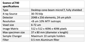
Scanco µCT40 scanner allows for visualization, measurement, and quantification of 3D structures
- Calibrated to measure density of mineralized specimens
- Contrast reagents can be used to visualize soft tissues, such as blood vessels
- This system creates a 3D model from a stack of 2D X-ray images taken around a single axis of rotation
- Resolution of 6 to 72 microns depending on sample size and scan parameters
- Specific trabecular or cortical analysis of long bones can be performed
- Equipped with an automatic sample changer that allows high throughput scanning
- Max specimen size 37 mm diameter x 80 mm length
Scanco uCT40 specifications
Pricing
Pricing is sample-dependent. Please contact the Core for a quote specific to your project.

MicroCT Core Facilities and Services
Advanced Microscopy Core Manager Jennifer Gundrum has over seven years of experience in advanced imaging services. Contact her for inquiries about services.


